

Gel Documentation
Gel Documentation Systems: Imaging, Analysis, and DNA Visualization Guide
Gel documentation systems are essential molecular biology tools used to visualize, capture, and analyze DNA, RNA, and protein samples after electrophoresis. This guide explains how gel imaging works, compares UV and blue-light systems, and helps you choose the right gel documentation system for your laboratory workflow.
What is Gel Documentation? →
Gel documentation (gel doc) refers to the process and instrumentation used to visualize, capture, and analyze nucleic acids (DNA or RNA) or proteins after electrophoresis. After samples are separated in an agarose or polyacrylamide gel, they are stained with fluorescent or intercalating dyes that allow the separated bands to be detected under specific wavelengths of light.
A gel documentation system typically consists of a light source (such as UV, blue light, or LED illumination), a dark imaging chamber, and a camera system that captures high-resolution images of the stained gel. These images are then used for qualitative and quantitative analysis of nucleic acid size, integrity, and abundance.
DNA visualization relies on fluorescent dyes that bind to nucleic acids. Common stains intercalate between DNA bases or bind within the minor groove, emitting fluorescence when excited by a specific wavelength. The resulting signal allows researchers to detect DNA fragments that would otherwise be invisible to the naked eye.
Traditional systems use ultraviolet (UV) transillumination, which provides high sensitivity but can cause DNA damage and poses safety concerns for users. Modern gel documentation systems increasingly use blue light or LED-based illumination, which reduces DNA damage and improves safety while maintaining high imaging quality for most routine molecular biology applications.
Gel documentation systems are widely used in cloning, PCR validation, restriction enzyme digestion analysis, CRISPR workflows, and RNA analysis. High-quality imaging is critical for accurately determining fragment size, verifying experimental success, and quantifying band intensity.
In addition to imaging, advanced gel documentation systems may include software for densitometry analysis, molecular weight estimation, and quantitative comparison between samples. These tools enable researchers to extract meaningful quantitative data from gel images, supporting downstream experimental design and publication-quality figure generation.
Overall, gel documentation is an essential step in molecular biology workflows that bridges electrophoretic separation and data analysis, enabling accurate visualization and interpretation of nucleic acid and protein samples.
Why Gel Documentation Is Essential in Molecular Biology →
After electrophoresis, DNA and RNA fragments are invisible without fluorescent or intercalating dyes. Gel documentation systems convert these molecular signals into digital images that allow researchers to verify experimental results, measure fragment sizes, and quantify nucleic acid abundance.
Accurate gel imaging is critical in cloning, PCR validation, restriction enzyme digestion analysis, CRISPR gene editing workflows, and RNA expression studies. Without proper imaging systems, experimental interpretation becomes subjective and non-reproducible.
How Gel Documentation Systems Work →
- Electrophoresis separation: DNA, RNA, or proteins are separated based on size in an agarose or polyacrylamide gel.
- Staining: Samples are stained using fluorescent or intercalating dyes that bind nucleic acids.
- Excitation: The gel is illuminated using UV, blue light, or LED excitation depending on system design.
- Emission detection: Fluorescent signals emitted by the stain are captured by a high-resolution camera system.
- Image analysis: Software is used to quantify band intensity, estimate molecular weight, and generate publication-ready images.
UV vs Blue Light vs LED Gel Documentation Systems →
| Feature | UV Systems | Blue Light Systems | LED Systems |
|---|---|---|---|
| DNA Damage Risk | High | Low | Low |
| Detection Sensitivity | Very High | High | Moderate–High |
| User Safety | UV exposure risk | Safe | Safe |
| DNA Integrity | Can cause fragmentation | Preserved | Preserved |
| Best Use Case | High-sensitivity detection | Cloning, PCR, routine workflows | General imaging workflows |
How to Choose the Right Gel Documentation System →
Selecting the appropriate gel documentation system depends on your application, sensitivity requirements, and safety preferences.
- For routine PCR and cloning: Blue light systems are recommended due to their balance of safety and sensitivity.
- For high-sensitivity detection: UV-based systems may provide stronger signal detection for low-abundance targets.
- For DNA integrity preservation: Blue light or LED systems are preferred to avoid UV-induced DNA damage.
- For imaging flexibility: Systems compatible with multiple dyes and excitation sources provide the greatest versatility.
Applications of Gel Documentation Systems →
- PCR product validation
- DNA cloning and restriction digestion analysis
- CRISPR genome editing verification
- RNA integrity and expression analysis
- Protein electrophoresis imaging
- Molecular weight estimation and quantification
Looking for an alternative DNA stain to Ethidium Bromide and don't know which to choose? Start here →
SafeView™ Classic (G108) is our recommended stain to start as it can be used as a direct substitute for Ethidium Bromide and plugged simply into existing workflows.
If a loading dye stain is desired, Safe-Green (G108) is recommended.
Our Safe-Red line up (G108-R and G680) are available if a red dye is desired.
Safer than Ethidium Bromide
As a leader in lab safety, abm has developed a system that eliminates these hazards:
- Low-Voltage LED Technology: Minimizes electric shock risks.
- Non-Carcinogenic Dyes: first and second-generation EtBr alternatives.
- UV-Free Illumination: Eliminates harmful UV exposure by using LED light.
Achieve superior, sensitive results with abm’s advanced lab-safe and risk-free gel documentation system.
| Product Name | Cat. No. | UV | Blue Light | Sensitivity (per band) | Size | Price |
|---|---|---|---|---|---|---|
| Gel Imager | ||||||
| SafeViewER™ Imager | E1001 | ✔ | N/A | 1 Unit | ||
| Loading Dyes | ||||||
| Safe-Green™ | G108-G | ✔ | ✔ | 0.2 – 0.6 ng | 1.0 ml | |
| Safe-Red™ | G108-R | ✔ | ✔ | 0.6 – 1.0 ng | 1.0 ml | |
| Gel Casting Dyes | ||||||
| SafeView™ Classic | G108 | ✔ | ✔ | 1.0 – 2.0 ng | 1.0 ml | |
| Safe-Red™ Gel | G680 | ✔ | ✔ | 0.6 – 1.0 ng | 1.0 ml | |
| Post Staining Dyes | ||||||
| Safe-Red™ Gel | G680 | ✔ | ✔ | 0.6 – 1.0 ng | 1.0 ml | |
| SafeView™ Classic | G108 | ✔ | ✔ | 1.0 – 2.0 ng | 1.0 ml | |
SafeView™ DNA Stains
SafeView™ products are a new class of fluorescent nucleic acid gel stains for the detection of double-stranded DNA, single-stranded DNA and RNA in agarose gel electrophoresis. Four formulations are available, including green or red fluorescence and gel-casting or loading dye formats, making them compatible with any experimental preference or setup!
Safe
Easy-to-use
Sensitive

Increased sensitivity compared to ethidium bromide
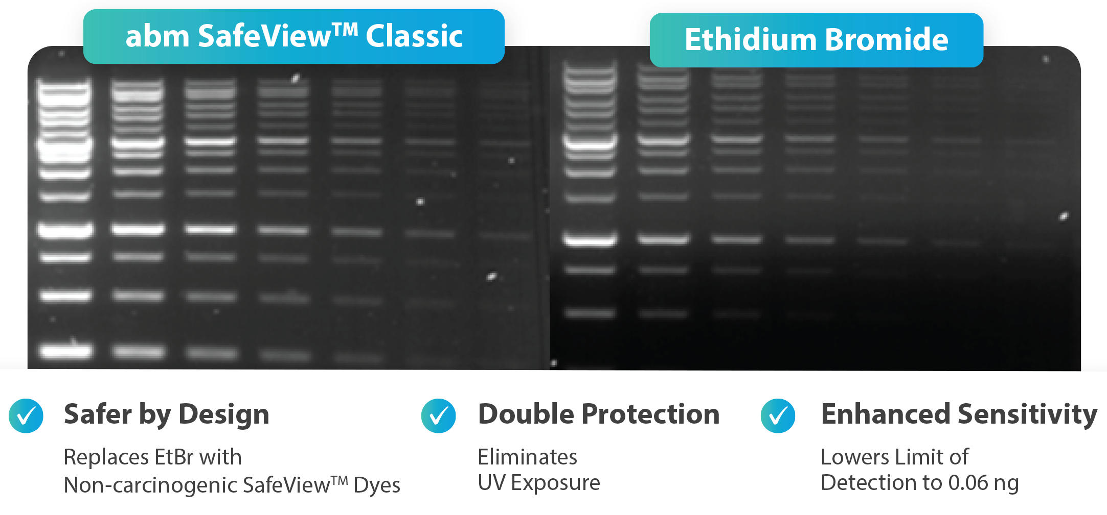
SafeView™ Classic has increased sensitivity compared to ethidium bromide. Two-fold serial dilution of abm’s 1kb Plus Opti-DNA Marker (Cat. No. G248) was visualized using SafeView™ Classic and ethidium bromide under the same conditions (1% agarose gel in TAE Buffer, SafeViewER™ Imager).
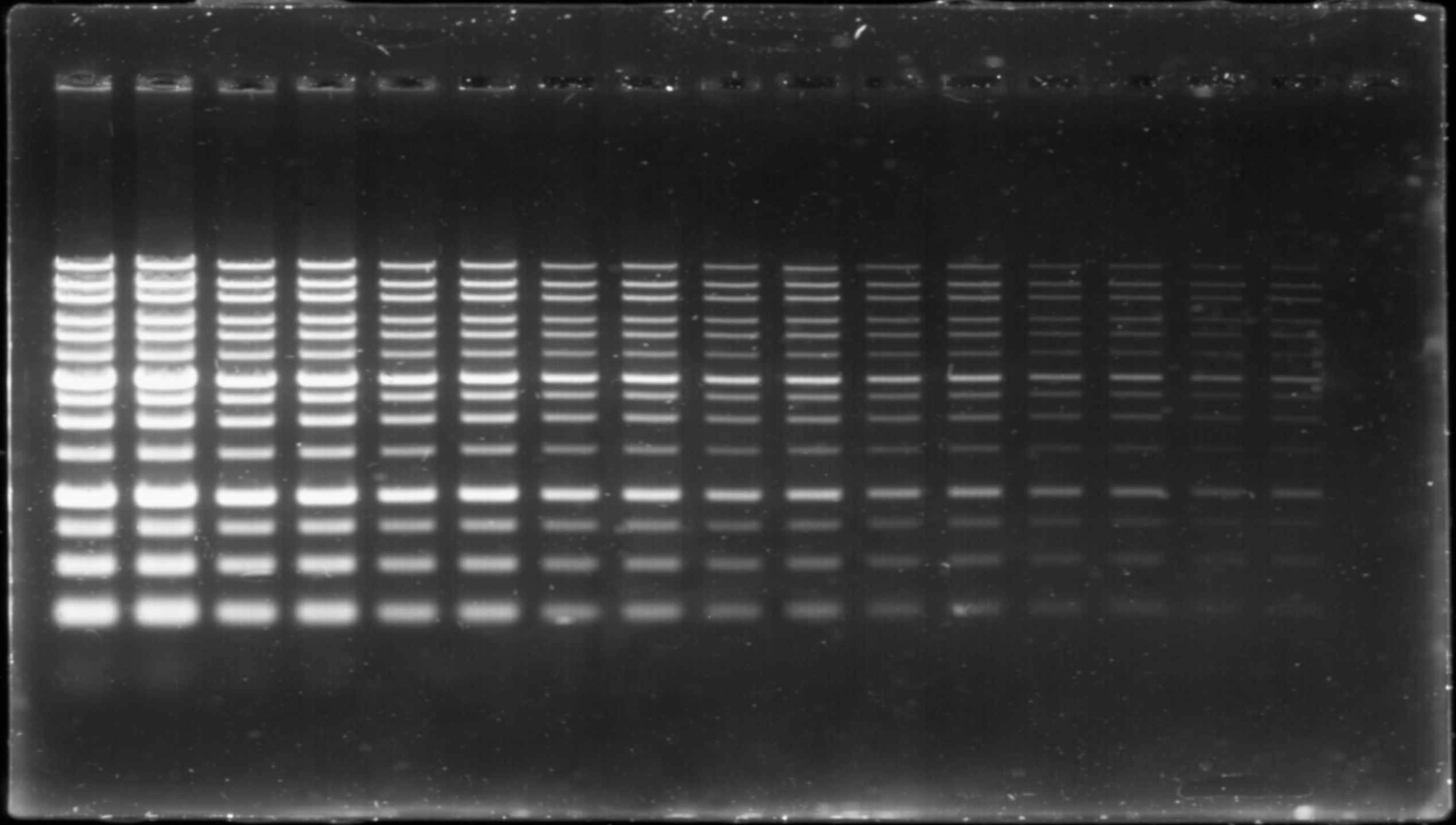
SafeView™ Classic is free of DNA migration issues that plague other dyes. Two-fold serial dilution of abm’s 1kb Plus Opti-DNA Marker (Cat. No. G248) was visualized using SafeView™ Classic on a 1% agarose gel in TAE Buffer with the SafeViewER™ Imager.
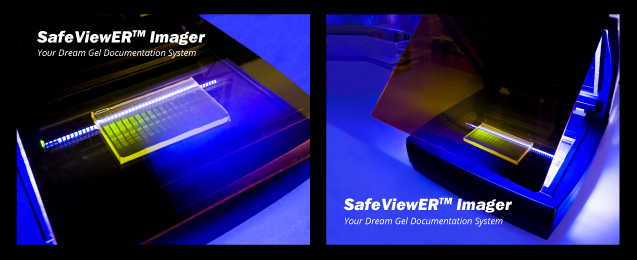
Meet the new SafeViewER™ Imager
- Safer by design: Replaces EtBr with non-carcinogenic SafeView™ dyes
- Double protection: Eliminates UV exposure
- Enhanced sensitivity: Lowers limit of detection to 0.06 ng
Click here to learn more about the SafeViewER™ Imager!
Frequently Asked Questions
What is the difference between G108, G108-G, G108-R, and G680 DNA Stains? →
The SafeView™ Classic (G108) and Safe-Red Gel (G680) series are gel casting dyes, meaning stains are added to the agarose gels as they are being made, while the Safe-Green (G108-G) and Safe-Red (G108-R) are gel loading dye, meaning they are added directly to the DNA sample as they are being loaded into the gel.
Which gel documentation system should I choose: G108, G108-G, G108-R, or G680? →
Our stains work with both UV and Blue LED Visualization. SafeViewER™ is especially great for use with SafeView™ Classic (G108).
What applications are your SafeViewER™ and SafeView™ Classic Stain used for? →
The system is used for routine gel imaging applications including PCR product verification, DNA cloning, restriction enzyme digestion analysis, and RNA quality assessment. It provides reliable imaging for standard molecular biology workflows.
Can the SafeView™ system be used for both DNA and RNA gels? →
Yes, SafeView™ Classic (G108) can be used for both DNA and RNA gel imaging.
Request a Free Sample
Simply select the free product sample(s) you would like to receive and we will get in touch with you to coordinate the delivery to your lab. Please note that sample availability is limited and honored on a "first come first served" basis and shipping charges may apply.