

SCCF1 Stably Expressing Luciferase Cell Line


Worried about losing your cells due to growth or thawing difficulties, or even a random freezer breakdown? Enjoy peace of mind knowing that you can be covered under abm's Cell Line Insurance.
Sale of this item is subjected to the completion of a Material Transfer Agreement (MTA) by the purchasing institution.
For for-profit organizations, please contact quotes@abmgood.com for pricing.
| Cat. No. | T6446 |
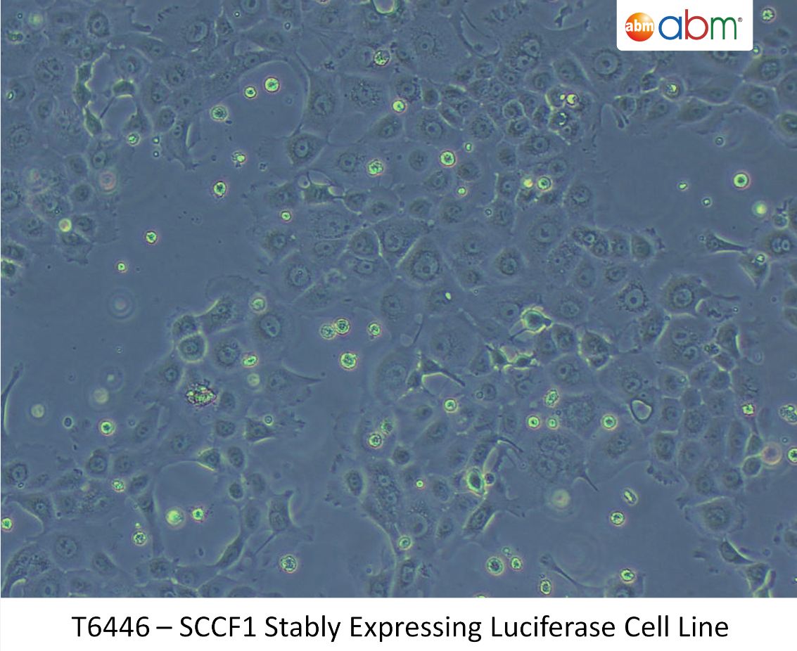
| Name | SCCF1 Stably Expressing Luciferase Cell Line |
| Description |
Developed from a laryngeal squamous cell carcinoma of a cat. The cells were stably transfected with a plasmid containing a luciferase-YFP fusion construct. SCCF1 cells can be used for experiments on genetic dysregulation in neoplastic keratinocytes of the feline oropharynx. Will form tumors in nude mice, but do not readily invade bone. These cells are a useful in vitro model for head and neck cancer. |
| Organism | Cat (Feline) |
| Tissue | Larynx |
| Donor History | Cat, Laryngeal squamous cell carcinoma |
| Growth Properties | Adherent, epithelial-like |
| Storage Condition | Vapor phase of liquid nitrogen, or below -130°C. |
| Shipping Conditions | Ship with dry ice. |
| Product Format | Frozen |
| Intended Use | This product is intended for laboratory research use only. It is not intended for any animal or human therapeutic use, any human or animal consumption, or any diagnostic use. |
| BioSafety | II |
| Certificate of Analysis | For batch-specific test results, refer to the applicable certificate of analysis that can be found at www.abmgood.com. |
| Growth Conditions |
For optimal cell culture, we recommend using PriCoat™ T25 Flasks
(G299)
or coating your preferred vessels with Applied Cell Extracellular Matrix
(G422).
PriGrow III (TM003) + 10% FBS(Regular*) + 1% Penicillin/Streptomycin Solution (G255), 37.0°C, 5% CO₂. *Do not heat-inactivate0.4 mg/ml Geneticin/G418 (G271) for selection. Note: Selection drugs should be added to the culture medium after the first passage to ensure cells have recovered from freeze-thaw conditions. |
| Unpacking and Storage Instructions |
1. Visually examine the packaging containers for signs of leakage or breakage. 2. Immediately transfer frozen cells from dry ice packaging to a temperature below -130°C, preferably in liquid nitrogen vapor phase storage, until ready for use. To ensure the highest level of viability, thaw the vial and initiate culture as soon as possible upon receipt. If continued storage is desired, the vial should only be stored below -130°C or in liquid nitrogen vapor phase. Do not store at -70°C, as it will result in loss of viability. |
| Thawing Protocol |
1. Thaw cells quickly in a 37°C water bath while agitating gently (maximum 2 minutes). The vial cap should be kept above the water level to minimize the risk of contamination. 2. Decontaminate the vial by spraying and wiping the exterior of the vial with 70% ethanol. From this point onwards, all operations should be strictly carried out inside a biological safety cabinet using aseptic conditions. 3. Transfer the cell suspension into a 15ml sterile conical tube containing 5ml of pre-warmed, complete growth media. Centrifuge cells at 125xg for 5-7 minutes. 4. Aspirate the supernatant without disturbing the cell pellet. Re-suspend the cell pellet in the recommended pre-warmed, complete growth media and dispense into a T25 culture flask. 5. Incubate the cells at the recommended conditions. |
| Subculture Protocol |
Volumes given below are for a T75 flask; proportionally increase or decrease the volume as required per culture vessel size. Subculture cells once the culture vessel is 80% confluent. 1. Aspirate the culture media, and add 2-3ml of pre-warmed 0.25% Trypsin-EDTA to the culture vessel. 2. Observe the cells under a microscope to confirm detachment (typically within 2-10 minutes). Cells that are difficult to detach can be put in 37°C, for several minutes to facilitate detachment. 3. Neutralize Trypsin-EDTA by adding an equal volume of the complete growth media into the culture vessel. 4. Transfer the culture suspension into a sterile centrifuge tube, and centrifuge at 125xg for 5 minutes. The actual centrifuge duration and speed may vary depending on the cell type. 5. Aspirate the supernatant, and re-suspend the pellet with pre-warmed fresh complete growth media. Add appropriate aliquots of the cell suspension to new culture vessels, as desired. 6. Incubate the cells at the recommended conditions. |
| Cryopreservation | We recommend using serum-free CryoGuard™ Freezing Media (TM078) or, if serum is preferred, Cryopreservation Medium (TM024). |
| Split Ratio | 1:5 |
| Expression |
Luciferase, YFP |
| Warranty | abm warrants that cell lines shall be viable upon initiation of culture for a period of thirty (30) days after shipment and that they shall meet the specifications on the applicable abm Material Product Information sheet, certificate of analysis, and/or catalog description. Such thirty (30) day period is referred to herein as the "Warranty Period”. |
| Disclaimer |
1. Sale of this item is subjected to the completion of a Material Transfer Agreement (MTA) by the purchasing individual/institution for each order. If you have any questions regarding this, please contact us at licensing@abmgood.com. 2. All test parameters provided in the CoA are conducted using abm's standardized culture system and procedures. The stated values may vary under the end-user's culture conditions. Please verify that the product is suitable for your studies by referencing published papers or ordering RNA (0.5 μg, Cat.# C207, $450.00) or cell lysate (100 μg, Cat.# C206, $600.00) to perform preliminary experiments, or alternatively use our Gene Expression Assay Service (Cat# C138). All sales are final. 3. We recommend live cell shipments for ease of cell transfer and this option can be requested at the time of ordering. Please note that the end-user will need to evaluate the feasibility of live cell shipment by taking into account the final destination's temperature variation and its geographical location. In addition, we thoroughly test our cell lines for freeze-thaw recovery. If frozen cells were received and not recovered in your lab under the exact, specified conditions (using recommended culture vessel, media, additional supplements, and atmospheric conditions), a live cell replacement is possible at a cost (plus shipping). 4. All of abm's cell biology products are for research use ONLY and NOT for therapeutic/diagnostic applications. abm is not liable for any repercussions arising from the use of its cell biology product(s) in therapeutic/diagnostic application(s). Please contact a technical service representative for more information. 5. abm makes no warranties or representations as to the accuracy of the information on this site. Citations from literature and provided for informational purposes only. abm does not warrant that such information has been shown to be accurate. 6. abm warrants that cell lines shall be viable upon initiation of culture for a period of thirty (30) days after shipment and that they shall meet the specifications on the applicable abm Material Product Information sheet, certificate of analysis, and/or catalog description. Such thirty (30) day period is referred to herein as the "Warranty Period." |
| Depositor | Ohio State University |
| QC |
1) Luminometer; 2) Flow cytometry |
| Application | Research Use Only. |
| Material Citation | If use of this material results in a scientific publication, please cite the material in the following manner: Applied Biological Materials Inc, Cat. No. T6446 |
|
How should I handle live cells once I receive them? |
|
|
Please refer to our Cell Handling and Thawing Guidelines for detailed instructions on receiving, thawing, and culturing live cells: |
| I want to make sure these cells express my gene of interest before I decide to buy the cell line. Can you provide a sample so this can be tested? | |
|
We do not perform extensive downstream characterization, or gene expression profiling of our cell lines. However, you can check the ‘References’ tab to see if a publication is available, or you can contact us to see if RNA or cell lysates are available for your cell line of interest. Please directly contact us for further information at quotes@abmgood.com.
|
| Does abm's PriGrow series medium contain antibiotics? | |
|
No, the medium does not contain antibiotics and needs to be supplemented by the end-user as desired.
|
| Do I need to use abm's media and Applied Cell Extracellular Matrix (G422) to culture my cells? | |
|
We strongly encourage using the recommended media and culture conditions described in the "Growth Conditions" section as they have been optimized for cell growth. Other supplier's media and coating conditions have not been tested.
|
| What is the difference between Applied Cell Extracellular Matrix (G422) and Collagen Coating? | |
|
The main component of our Applied Cell Extracellular Matrix (G422) is type I collagen specifically. For more information on abm's Applied Cell Extracellular Matrix please visit: http://www.abmgood.com/Applied-Cell-Extracellular-Matrix-G422.html.
|
| Do I have to use T25 ECM-coated flasks for growing the cells? | |
|
We strongly recommend thawing cells in the T25 flasks specified on the cell-specific product page to ensure optimal post-thaw recovery of the frozen cells before testing other culture vessels.
|
| If I want to plate these cells to multi-well plates (e.g. 96 well plates) or dishes, how should I prepare the plates? | |
| What percentage of Trypsin-EDTA should be used to subculture cells? Can I use Trypsin-EDTA containing phenol red? | |
|
The standard concentration used for subculturing is 0.25% Trypsin-EDTA, available at abm (Cat. No. TM050). Yes, Trypsin-EDTA containing phenol red can be used as it does not interfere with trypsinization.
|
| How often do I need to change the media? | |
|
Media should be changed every 2-3 days or as specified within the recommended growth conditions.
|
| Why are these cells classified as biosafety level II? | |
|
We follow the CDC-NIH recommendations that all mammalian sourced products should be handled at the Biological Safety Level 2 to minimize exposure of potentially infectious products. This information can be found in 'Biosafety in Microbiological and Biomedical Laboratories' (1999). Your institution's Safety Officer or Technical Services will be able to make the call as to whether BioSafety Level I is possible with these cells at your site, if desired.
|
| How long can I store frozen vials? | |
|
Cells that are properly frozen using an effective cryoprotective agent can be stored in liquid nitrogen vapor phase (or below -130°C) indefinitely without affecting viability.
|
| What is the recommended storage temperature for cells? | |
|
In general, if you received:
|
| Can I substitute heat-inactivated FBS with FBS or vice versa? | |
|
FBS and heat-inactivated FBS are different in their composition; they cannot be substituted for one another.
|
| My cells are not detaching, what method do you recommend to trypsinize the cells? | |
|
| How are live cells shipped? | |
|
Live cells are shipped as a live culture in a T12.5 flask. Cells are seeded at an optimal confluency to avoid metabolic waste build up during transport. In addition, we thoroughly test our cell lines for functional freeze-thaw recovery. For more information on how to handle live cells, please view our detailed instructions here (https://www.abmgood.com/uploads/document/Cell_Handling_Instructions_Upon_Arrival_150623.pdf).
|
| Why are cells not attaching and forming clumps after subculturing with trypsin? | |
|
Cells form clumps and display attachment issues when the trypsinization agent is not effectively neutralized. To completely neutralize the trypsin agent, add an equal amount of complete growth media prior to centrifugation. Subtle changes in culture conditions, particularly in pH and the quality of serum used in the growth medium, may also affect the amount of clumping exhibited by the cells.
|
| Why are my cells forming clumps after plating? | |
|
It is important to ensure cell clumps are broken up through gentle re-suspension by pipetting up and down several times after pelleting cells by centrifugation. This will ensure a homogenous single cell suspension for even plating.
|
| Do I need to add the selection drug to the complete growth medium? | |
|
We recommend maintaining selection pressure using the drug concentration specified in the Growth Conditions section.
|
| Where can I find the CoA for my product? | |
|
Certificates of analysis can be found here (https://www.abmgood.com/CoA-Library.html).
|
|
What is your warranty or return policy? |
|
|
Our warranty and return policy is outlined in abm’s Terms and Conditions, including details on product quality, limitations, and claims. For additional questions, our Order team is happy to assist and can be reached at order@abmgood.com. |
|
How many times can cells divide? |
|
|
The number of times cells can divide depends on the cell type:
|
|
What is the difference between spontaneous immortalization and tumor-derived cells? |
|
|
Spontaneously immortalized cells arise when normal cells acquire the ability to proliferate indefinitely in culture without intentional genetic manipulation. These cells are not derived from tumors but may undergo genetic or phenotypic changes during immortalization.
Tumor-derived cells are established directly from cancerous tissue and typically retain characteristics associated with malignancy, such as altered growth control and genetic instability. |
|
Do I need Applied Cell Extracellular Matrix (G422) if I am using PriCoat™ flasks? |
|
|
Please refer to the Growth Conditions section of your specific product page. This section will indicate whether Applied Cell Extracellular Matrix (G422), PriCoat™ flasks, or both are recommended for optimal cell attachment and growth, as requirements may vary by cell type. |
-
Tannehill-Gregg, S., Kergosien, E., & Rosol, T. J. (2001). Feline head and neck squamous cell carcinoma cell line: characterization, production of parathyroid hormone-related protein, and regulation by transforming growth factor-beta. In vitro cellular & developmental biology. Animal, 37(10), 676–683. https://doi.org/10.1290/1071-2690(2001)037
Tannehill-Gregg, S. H., Levine, A. L., & Rosol, T. J. (2006). Feline head and neck squamous cell carcinoma: a natural model for the human disease and development of a mouse model. Veterinary and comparative oncology, 4(2), 84–97. https://doi.org/10.1111/j.1476-5810.2006.00096.x
Heller, D. A., Fan, T. M., de Lorimier, L. P., Charney, S. C., Barger, A. M., Tannehill-Gregg, S. H., Rosol, T. J., & Wallig, M. A. (2007). In vitro cyclooxygenase-2 protein expression and enzymatic activity in neoplastic cells. Journal of veterinary internal medicine, 21(5), 1048–1055. https://doi.org/10.1892/0891-6640(2007)21[1048:ivcpea]2.0.co;2
Wypij, J. M., Fan, T. M., Fredrickson, R. L., Barger, A. M., de Lorimier, L. P., & Charney, S. C. (2008). In vivo and in vitro efficacy of zoledronate for treating oral squamous cell carcinoma in cats. Journal of veterinary internal medicine, 22(1), 158–163. https://doi.org/10.1111/j.1939-1676.2007.0010.x
Bergkvist, G. T., Argyle, D. J., Morrison, L., MacIntyre, N., Hayes, A., & Yool, D. A. (2011). Expression of epidermal growth factor receptor (EGFR) and Ki67 in feline oral squamous cell carcinomas (FOSCC). Veterinary and comparative oncology, 9(2), 106–117. https://doi.org/10.1111/j.1476-5829.2010.00239.x
Bergkvist, G. T., Argyle, D. J., Pang, L. Y., Muirhead, R., & Yool, D. A. (2011). Studies on the inhibition of feline EGFR in squamous cell carcinoma: enhancement of radiosensitivity and rescue of resistance to small molecule inhibitors. Cancer biology & therapy, 11(11), 927–937. https://doi.org/10.4161/cbt.11.11.15525
Wakshlag, J. J., Peters-Kennedy, J., Bushey, J. J., & Loftus, J. P. (2011). 5-lipoxygenase expression and tepoxalin-induced cell death in squamous cell carcinomas in cats. American journal of veterinary research, 72(10), 1369–1377. https://doi.org/10.2460/ajvr.72.10.1369
E. Balkman, C. , L. Gieger, T. , M. Zgola, M. , D. Lewis, L. and C. McEntee, M. (2012) In Vitro Characterization of Docetaxel as a Radiosensitizer in Canine and Feline Cancer Cell Lines. Open Journal of Veterinary Medicine, 2, 285-292. doi: 10.4236/ojvm.2012.24045.
Martin, C. K., Dirksen, W. P., Shu, S. T., Werbeck, J. L., Thudi, N. K., Yamaguchi, M., Wolfe, T. D., Heller, K. N., & Rosol, T. J. (2012). Characterization of bone resorption in novel in vitro and in vivo models of oral squamous cell carcinoma. Oral oncology, 48(6), 491–499. https://doi.org/10.1016/j.oraloncology.2011.12.012
Pang, L. Y., Bergkvist, G. T., Cervantes-Arias, A., Yool, D. A., Muirhead, R., & Argyle, D. J. (2012). Identification of tumour initiating cells in feline head and neck squamous cell carcinoma and evidence for gefitinib induced epithelial to mesenchymal transition. Veterinary journal (London, England : 1997), 193(1), 46–52. https://doi.org/10.1016/j.tvjl.2012.01.009
Jackie M. Wypij et al., Surrogate biomarkers interleukin-6 and interleukin-10 in a spontaneous feline model of head and neck squamous cell carcinoma (HNSCC).. JCO 32, e17056-e17056(2014).doi:10.1200/jco.2014.32.15_suppl.e17056
Brown, M. E., Bear, M. D., Rosol, T. J., Premanandan, C., Kisseberth, W. C., & London, C. A. (2015). Characterization of STAT3 expression, signaling and inhibition in feline oral squamous cell carcinoma. BMC veterinary research, 11, 206. https://doi.org/10.1186/s12917-015-0505-7
Parviainen, S., Autio, K., Vähä-Koskela, M., Guse, K., Pesonen, S., Rosol, T. J., Zhao, F., & Hemminki, A. (2015). Incomplete but infectious vaccinia virions are produced in the absence of oncolysis in feline SCCF1 cells. PloS one, 10(3), e0120496. https://doi.org/10.1371/journal.pone.0120496
Supsavhad, W., Dirksen, W. P., Hildreth, B. E., & Rosol, T. J. (2016). p16, pRb, and p53 in Feline Oral Squamous Cell Carcinoma. Veterinary sciences, 3(3), 18. https://doi.org/10.3390/vetsci3030018
Supsavhad, W., Dirksen, W. P., Martin, C. K., & Rosol, T. J. (2016). Animal models of head and neck squamous cell carcinoma. Veterinary journal (London, England : 1997), 210, 7–16. https://doi.org/10.1016/j.tvjl.2015.11.006
Cannon, C. M., Trembley, J. H., Kren, B. T., Unger, G. M., O'Sullivan, M. G., Cornax, I., Modiano, J. F., & Ahmed, K. (2017). Evaluation of protein kinase CK2 as a therapeutic target for squamous cell carcinoma of cats. American journal of veterinary research, 78(8), 946–953. https://doi.org/10.2460/ajvr.78.8.946
Cannon, C. M., Trembley, J. H., Kren, B. T., Unger, G. M., O'Sullivan, M. G., Cornax, I., Modiano, J. F., & Ahmed, K. (2017). Therapeutic Targeting of Protein Kinase CK2 Gene Expression in Feline Oral Squamous Cell Carcinoma: A Naturally Occurring Large-Animal Model of Head and Neck Cancer. Human gene therapy. Clinical development, 28(2), 80–86. https://doi.org/10.1089/humc.2017.008
Trembley, J. H., Kren, B. T., Abedin, M. J., Vogel, R. I., Cannon, C. M., Unger, G. M., & Ahmed, K. (2017). CK2 Molecular Targeting-Tumor Cell-Specific Delivery of RNAi in Various Models of Cancer. Pharmaceuticals (Basel, Switzerland), 10(1), 25. https://doi.org/10.3390/ph10010025
Nasry, W. H. S., Wang, H., Jones, K., Dirksen, W. P., Rosol, T. J., Rodriguez-Lecompte, J. C., & Martin, C. K. (2018). CD147 and Cyclooxygenase Expression in Feline Oral Squamous Cell Carcinoma. Veterinary sciences, 5(3), 72. https://doi.org/10.3390/vetsci5030072
Piegols, H. J., Takada, M., Parys, M., Dexheimer, T., & Yuzbasiyan-Gurkan, V. (2018). Investigation of novel chemotherapeutics for feline oral squamous cell carcinoma. Oncotarget, 9(69), 33098–33109. https://doi.org/10.18632/oncotarget.26006
Harris, K., Gelberg, H. B., Kiupel, M., & Helfand, S. C. (2019). Immunohistochemical Features of Epithelial-Mesenchymal Transition in Feline Oral Squamous Cell Carcinoma. Veterinary pathology, 56(6), 826–839. https://doi.org/10.1177/0300985819859873
Renzi, A., De Bonis, P., Morandi, L., Lenzi, J., Tinto, D., Rigillo, A., Bettini, G., Bellei, E., & Sabattini, S. (2019). Prevalence of p53 dysregulations in feline oral squamous cell carcinoma and non-neoplastic oral mucosa. PloS one, 14(4), e0215621. https://doi.org/10.1371/journal.pone.0215621
Borlle, L., Dergham, A., Wund, Z., Zumbo, B., Southard, T., & Hume, K. R. (2019). Salinomycin decreases feline sarcoma and carcinoma cell viability when combined with doxorubicin. BMC veterinary research, 15(1), 36. https://doi.org/10.1186/s12917-019-1780-5